Dr Sarah Bohndiek (Biological and Soft Systems) and Professor Mete Atatüre and Dr Helena Knowles (Atomic, Mesoscopic and Optical Physics)
This project combines two novel sensing approaches to answer the following open questions in oxidative stress biology:
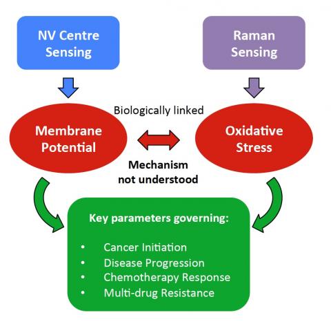
1. How do cells adapt their plasma membrane chemistry in response to oxidative stress?
2. Do these changes help to mediate the common emergence of multidrug resistance in disease, in particular, lung cancer?
The multidisciplinary connections between physical (blue) and chemical (purple) sensing, biochemistry (red), and biomedical applications (green) are required to answer these questions. Collaboration between Prof. Atature (NV sensing) and Dr Bohndiek (Raman sensing) brings together two crucial technologies (blue, purple), in a wet lab environment, which provides a unique opportunity to advance single cell biochemistry. Our goal is to create a system that facilitates simultaneous measurement of physiological parameters with these tools in living cells, to study their impact on drug response and resistance in lung cancer.
Project Completed
This project combines sensing of the intracellular environment (based on diamond spins) with sensing of the cellular biochemistry (based on Raman spectroscopy) to evaluate oxidative stress conditions in cancer cells. Oxidative stress, referring to an imbalance of damaging reactive oxygen species (ROS) and compensating antioxidants in cells, is a key marker in the incidence and progression of several diseases, including cancer. Current methods for evaluation of oxidative stress take bulk measurements of millions of cells, so have limited sensitivity to the heterogeneity of cellular responses. Through the course of this project, we created a unique microscope that enables for the first time non-toxic, real-time measurements of the chemical and environmental response to oxidative stress at the single cell level. We have thoroughly characterised the technical performance of this microscope, and applied it to evaluate the impact of different fluid environments on the fluorescence response of nanodiamonds. We have also quantitatively determined the biological impact of nanodiamonds in cultured cancer cells (see Figure). We are currently progressing towards measurement of oxidative stress in live cells, targeting cancer cells with different chemotherapy drugs. Ultimately, making such measurements could open opportunities for early identification of drug response and resistance, optimising treatment for cancer patients.
Figure: Nanodiamonds are endocytozed into breast cancer cells. Blue is NucBlue nuclear stain, orange is WGA-AF488 cell membrane cell, white shows the nanodiamonds (both individual and as aggregates) inside MDA-MB-231 breast cancer cells in vitro. This image is taken after 48 hours under a diamond exposure of 1ug/ml.